
A mix of new technology and old standbys is helping prevent and diagnose vision problems.
Technology has really improved the eye exam experience. There are still low tech aspects to even the best exams, but for the most part, eye exams have become more efficient and more effective.
Here’s what you need to know:
- Be prepared for questions. Eye exams typically start with a brief family and medical history – just tell us how much you know! For example if someone in your family used eye drops but you don’t know what for, this information is a clue. You should disclose pre-existing health conditions, such as diabetes or high blood pressure, and any medications that you take.
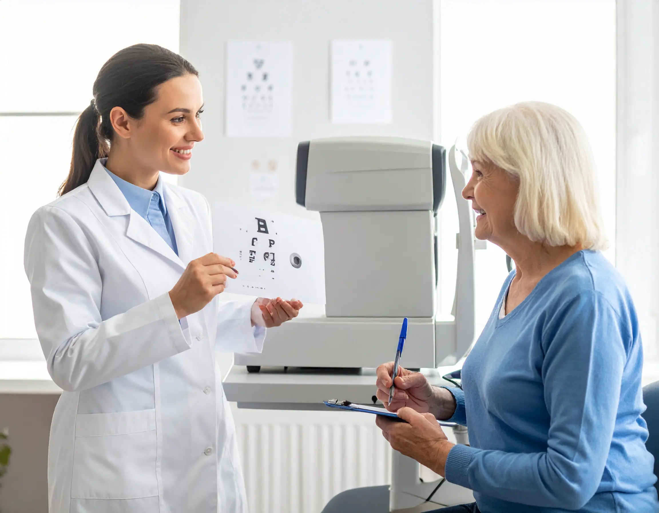
- Yes, the eye chart is still a thing! Our eye docs use a high tech computer version of the Snellen chart on a wall mounted flat screen while the doctor changes he picture with a remote. Same concept as ‘what is the smallest line that you can read?’
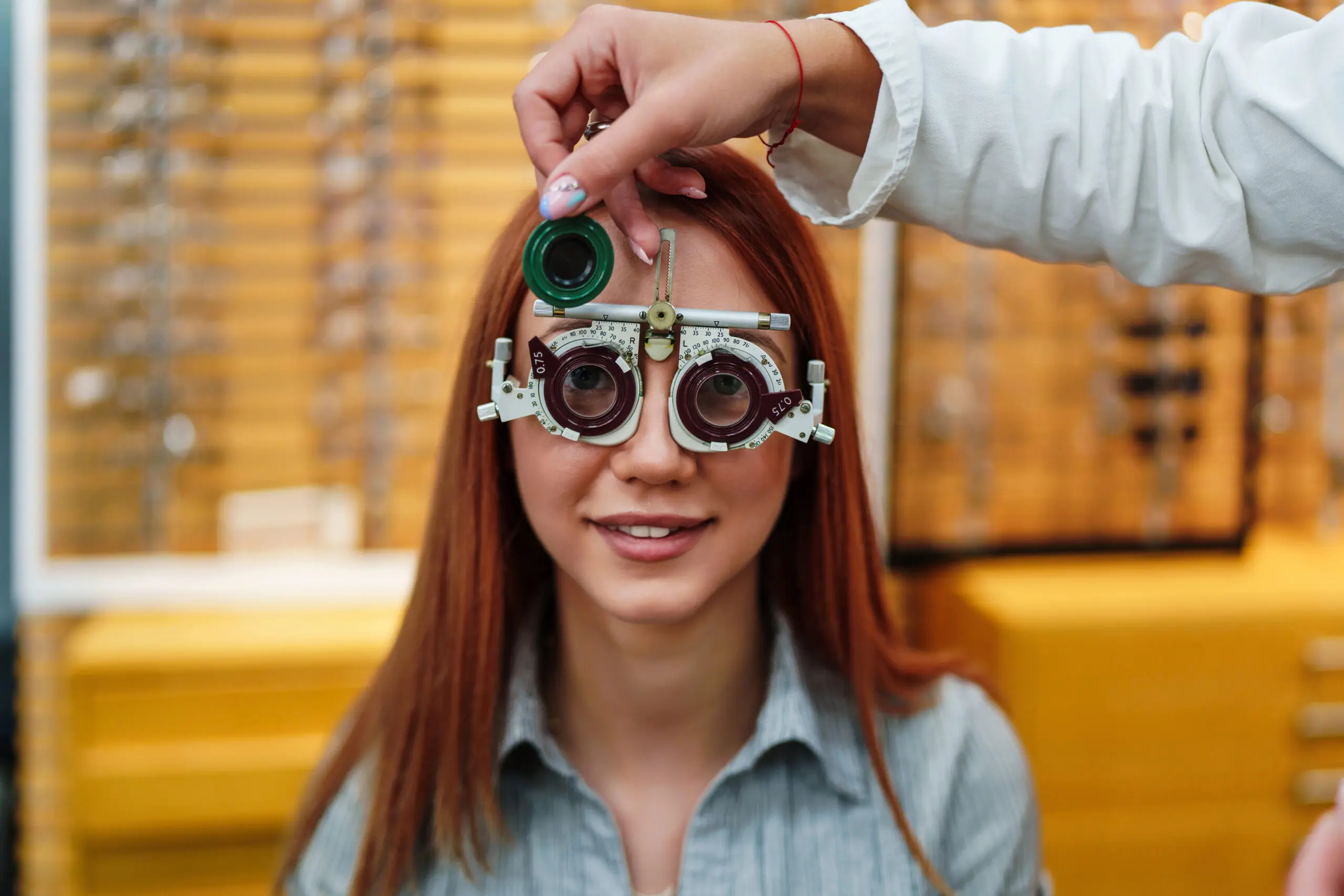
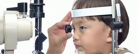
- A newer way to check your prescription. The old school retinoscope, the instrument that the doctor used to shine a light in your eyes, measured how light changes as it enters your eye. That’s called the ‘refractive error’, and it helps determine prescriptions for glasses or contact lenses. Now the doctor uses an autorefractor which does the same thing more objectively! Getting your correct prescription happens now more quickly. There’s still some fine tuning that the doctor uses – a series of slightly different lenses in front of your eyes to check which one helps you see better~~ “1 or 2?’
- Don’t fear the air puff! Increased eye pressure is a red flag for glaucoma, a disease that damages the optic nerve and can lead to vision loss. During most eye exams, our docs use a tonometer – that’s when you feel the quick puff of air – to measure the pressure in your eye. Guess what? It’s painless and you’ll be glad that you did it.
- Our docs can get a closer look with digital images. With our OCT (optical coherence tomography), your doctor can take digital images of the retina’s layers, mapping and measuring thickness. The retina, at the back of the eyeball, contains cells that are sensitive to light and that trigger nerve impulses that pass through the optic nerve to the brain, allowing for vision. The images are so good that our docs are almost looking at a biopsy of the tissue, and the measurements help determine if you have a healthy retina or serve as a tip off for conditions such as diabetic eye or age related macular degeneration, a leading cause of blindness.
- Dilation is a maybe……. Your doctor may still need to dilate your eye using special eye drops to enlarge pupils. Do not skip this step if it’s recommended! Yes, it is a hassle because it takes extra time and blurs your vision for an hour or more, but it still is the only way to get a true picture of the back of the eye, allowing doctors to look for indications of trouble. It can also help doctors detect signs of health problems like diabetes, high cholesterol, hypertension or thyroid disease, long before you notice symptoms. Should you decide against it, then be sure to choose to have the Optomap which allows the doctor to see 83% of the eye.
- Your exam may be free. While eye exams are reasonably priced, especially if you have vision coverage, regular health insurance does cover some checkups, especially if you have a health condition like diabetes.
- Contact Lens Eye Exam. Be sure to let the team know if you want to try contacts – it takes the doc a little more time to figure out the best contacts for you to start with and then to learn contact lens protocol.